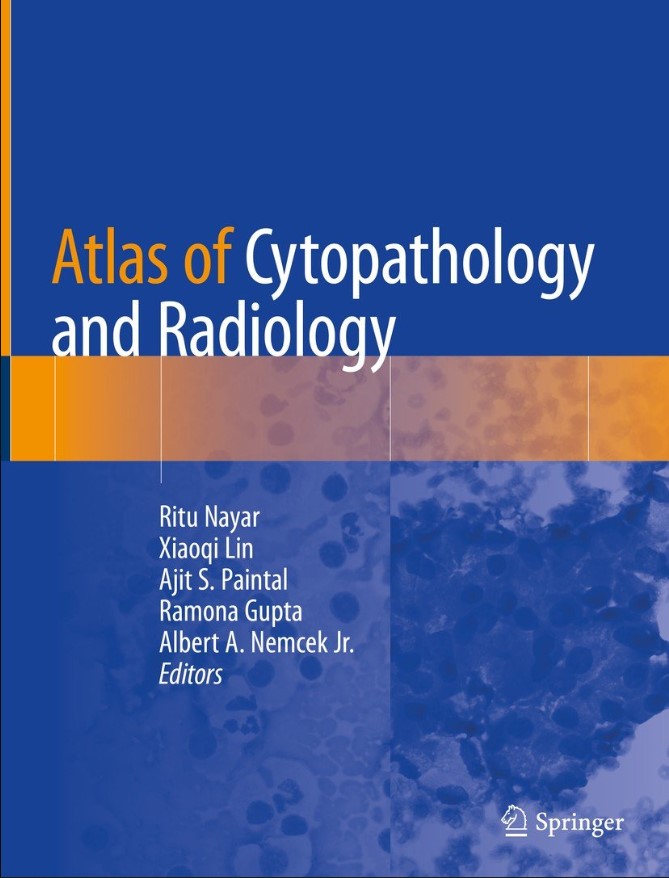
Atlas of Cytopathology and Radiology
The aim of this atlas is to guide pathologists and radiologists in the accurate triage and diagnosis of deep seated mass lesions biopsied under ultrasound, CT Scan or fluoroscopic guidance.
Fine needle aspiration cytology has become the foremost diagnostic modality in recent years for the diagnosis of mass lesions, including primary and recurrent neoplasms and masses of non-neoplastic and infectious etiology. An essential requirement in the accurate diagnosis of these masses is the correlation of cytomorphology with the radiological findings and adequate triage of acquired material during the biopsy procedure. The cytologic appearance (fine needle aspiration smears, touch preparations, cell blocks and core biopsy), gross surgical resected specimen (where available on follow-up) and imaging findings will be illustrated to provide a complete pathologic-radiologic correlation of the entities discussed. Collection methods and correlation with ancillary studies such as flow cytometry, microbiologic cultures, cytogenetics and immunohistochemistry will be described. The importance of specimen type and cytologic and radiologic techniques will be emphasized.
- Pages : 251 pages
- Publisher: Springer; 1st ed. 2020 edition (November 5, 2019)
- Language: English
- ISBN-10: 3030247546
- ISBN-13: 978-3030247546
- Product Dimensions: 6.9 x 9.8 inches


